August 11, 2022
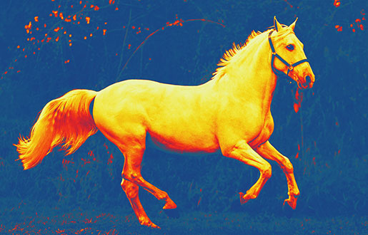
Strength, agility, grace, intelligence, nobility, will to be free, boundless loyalty, irrepressible energy, beauty, vulnerability, and vulnerability simultaneously. This organically combines a graceful animal covered with legends, glory, ancient myths, and gratuitous loyalty to man. It is a horse. Every nation, in its way, shows the uniqueness and splendor of this animal, improving and perfecting for centuries breeds of horses, distinguishing them by their colors and exterior, keeping the history of their origin, and noting their merits the society. In the Arab world, the horse is believed to be a gift from God, brought down from his hands and the wind to the earth. The horse has always been the most valuable animal in the family, the breadwinner in all times, the friend and warrior in times of war, and the healer and comforter in hopelessness and turmoil. Today, the horse is not only for work and sporting events but, as scientists have proven, it has a positive effect on the human nervous system, due to which animals are used in the treatment of many diseases and rehabilitation. The horse is a loyal animal and has always tried to show his loyalty to his master. And what can we do in return for this animal, helping him maintain his health? After all, there are plenty of problems, some hidden from our view "under the saddle." According to some statistics, when examining sport and hobby horses, more than seventy percent of horses have sore backs due to improperly sized saddles alone. In addition, torn ligaments, damaged muscles, tendons, dislocated spinal cartilage, joint problems, and more. Everything is just like in humans. Only the animal can't tell about it. Detecting these diseases in their early stages is the main task that infrared thermal imaging can help with.
How it works
A little history. In 1956 the Canadian surgeon Lohsen used a night vision device for the first time in clinical practice to diagnose the early stages of cancer. Until then, it was used exclusively for military purposes. Further research on this technology in early breast cancer diagnosis in women showed stunning results - almost seventy percent of positive diagnoses. It was a breakthrough. So what is it, and how does it work in veterinary medicine? Thermal imaging diagnostics is, conventionally speaking, a scanner that takes information about infrared radiation from an animal's body and transmits that data to a computer for processing through a system of focusing mirrors. The device that does this thermal imaging of the temperature distribution on the surface under study is a thermal imager. Its value lies in the "sees" invisible infrared thermal radiation.
Moreover, even directly in the thermal spectrum, it finds differences in temperature modes, showing them on the monitor as a color picture in the form of a stretched mosaic, where from pink to red - the scale of warm readings, and blue and black gradation is responsible for the cold areas. This is a critical point since all horse diseases and humans are within us. For example, the animal's knee joint is inflamed. Nothing yet portends trouble. The center of inflammation enlarges, and the horse's body sends us signals, but we do not notice them until our friend begins to limp and stop in pain. And yet the disease could have been prevented by doing a completely painless, no-contact examination of the animal for inflammation and, with a proper diagnosis, taking the necessary treatment measures. The study of thermal images is called thermography. Since the physical component of the living body, including inflamed areas, emits infrared vision, the temperature and radiation readings change accordingly. Thermography "encompasses" the object under study and not simply scans it by thermal parameters but "decomposes" this object into individual thermal points. In doing so, it analyzes the thermal readings and diagnoses and indicates the location of the inflammation. This is often the only method that helps identify various diseases in different parts of the animal's body.
What is it used for?
Pathologies that yesterday were detected when the disease was irreversible. Today, thanks to thermal vision, they are diagnosed in the early stages, sometimes long before the apparent manifestations of the disease. An infrared camera for equine examination gives the veterinarian a high-resolution image of the area under review with a detailed report indicating the problem areas. This makes it possible to see inflammation much earlier than it manifests itself. The list of problems is quite broad. These are joint diseases, arthritis, arthrosis, osteoarthritis, detection of hoof abscesses,
injuries to muscles, ligaments, tendons, and other inflammations that can lead to severe consequences. The tomograph, among other things, sees the inflamed place at the stage of disease development, when it is still possible to start successful medical treatment and remove the problem in time. Moreover, the range of action is comprehensive. Urology, traumatology,
pharmacology, oncology, and others. Measuring the temperature in animals allows you to think about when it rises. High temperature is a "bell" of the problem. And in what form it will appear is a matter of time. It may be the banal flu, but there may be unexpected options. Skin diseases, scabies, shingles, moss, chorioptosis, rheumatoid inflammatory processes. Clearly, in these cases, the thermometer only informed that it is necessary to connect the infrared vision. However, keep in mind that thermal imaging cameras, for all their capabilities, are not medical equipment, and you should not expect them to be able to detect a horse or other animal's illness one hundred percent of the time. Learn a simple truth: consider thermal imaging an effective primary preclinical inspection tool.
Conditions diagnosed with thermal imaging
Thermal imaging is the only method of non-contact animal diagnosis. Thermal imaging cameras in a particular range very well pick up and read the radiation emitted by the animal's body while being able to pick out the areas that signal a problem. In this situation, there is the possibility of prediction. In other words, the process becomes manageable, the situation understandable. Remote thermography is a safe and non-contact way to "scan" the animal's body from half a meter distance. The resulting thermographic thermal pattern is evaluated according to visual and quantitative criteria. In the first variant, the places with problematic temperature differences, their locations with analysis of hot spots, and approximate evaluation of quantitative radiation are considered and analyzed. In this case, it should be understood that achieving a correct temperature measurement will not be possible. Because of the small size of the surveyed area, the results are uncertain. The quantitative method is more promising. It is a modern technology used for prevention, evaluative analysis during research, and recommendations and suggestions for effective tomograph operation.
Finally, a few words about the prospects for improving thermal imaging: we are talking about improving the quality of images and obtaining images of problematic areas with detailing and automation of examinations. And also improvement of thermal imaging methods for studying different kinds of animal diseases and developing equipment working in the long-wave spectral range for fixation of maximum thermal radiation.